Hi all,
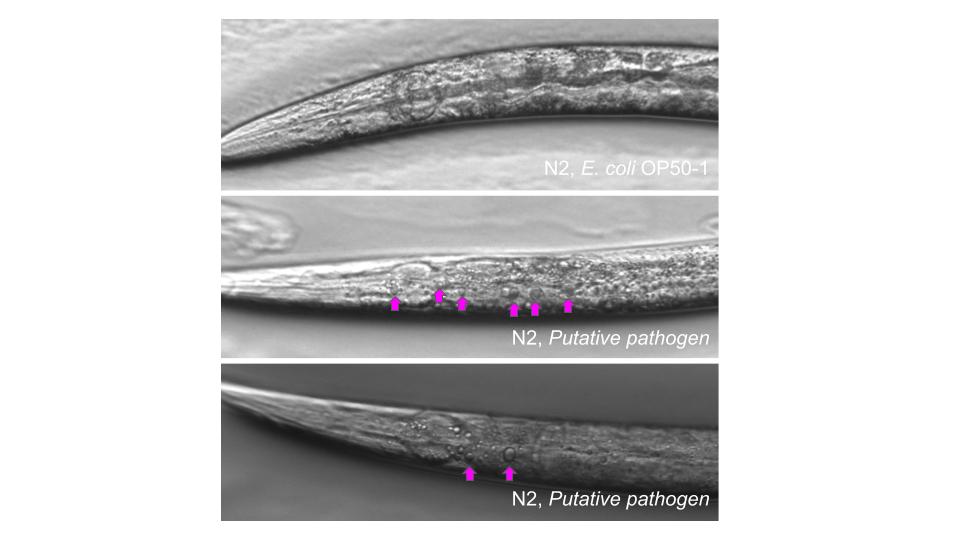
I’m exposing worms to a putative pathogen and I reproducibly find what look like large spheres showing up in animals. They look a little like supersized lipid droplets (Genetic Screen for Mutants with Supersized Lipid Droplets in Caenorhabditis elegans | G3 Genes|Genomes|Genetics | Oxford Academic) or maybe like vacuoles (Cells | Free Full-Text | Caenorhabditis elegans Deficient in DOT-1.1 Exhibit Increases in H3K9me2 at Enhancer and Certain RNAi-Regulated Regions).
I understand that there are molecular approaches I can take to figure this out, but wanted to know if this is something some of you may have experience with.
Here’s a picture!
Thanks,
Kris
These spheres look possibly similar to the vacuoles induced by lethal infection of C. elegans by Leucobacter Verde2 – see Figure 4E,F in Hodgkin et al. 2013
Curr Biol. 2013 Nov 4;23(21):2157-61. doi: 10.1016/j.cub.2013.08.060. Epub 2013 Oct 24.
We still don’t know how these vacuoles form; hasn’t been further investigated.
Thank you so much! They are very intriguing. I’ll take a look at your paper. Cheers!
While looking at pond snail (Lymnaea) hemolymph, my student found a spherical vesicle moving slowly around… clearly not drifting but somehow being driven. I think it must have been tethered to a swimming bacterium. Maybe the same kind of thing? I suggest you bathe your worms with diOC16, which will light up all lipids: a confocal view will tell you whether you’re looking at a solid fat-ball or a membrane-enclosed structure with aqueous insides.
Thanks for the response…I’ve since stained them with Oil-Red-O and they’re lipids! Best, Kris